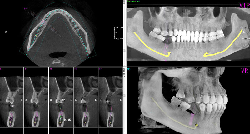
CN-Pano3, CN-Pano3C OPG X-Ray Machine are a cone beam volumetric tomography and panoramic X-ray device used for dental head and neck applications. CN-Pano3, CN-Pano3C using cone beam, a computer can reconstruct a three-dimensional image of the site after a single scan on oral maxillofacial region. It can clearly display three-dimensional body maxillofacial anatomy and achieve any level of cutting, accurate measurement of distance, surface area, volume and contour outline, solve the two-dimensional flat plate technology inherent in the image overlay, distortion and other issues. Compared with conventional CT, oral CT system is low cost, small footprint, low-dose, short shooting time, high resolution, easy operation.
MODEL | PANOROMA | PART CBCT | COMPUTER & 3D SOFTWARE (DCT VIEWER) | CEPHALOMETRIC |
CN-Pano3 | √ YES | √ YES | √ YES | × NO |
CN-Pano3C | √ YES | √ YES | √ YES | √ YES |
FEATURES
1>Mixed-mode radiation: Low-dose pulse radiation;
2>High Voxel Reconstruction: FOV 3X4 70µm ;
3>Special algorithm for Metal Artifact Reduction;
4>The most advanced technology for X-ray conversion, cesium iodide directly filmed on the surface of the sensor, to ensure ultra-high-definition images.
5>Software runs perfectly with PACS used in china hospitals. Optional software customized to specific use with completely independent intellectual property rights;
6>Flexible configuration and upgrades values for money, and short term for ROI;
7>A global community of after sales network

APPLICATION
With FOV 12*8, it can be applied in normal dental disease diagnosis, full mouth reconstruction Oral Surgery, teeth implant design etc.

TECHNICAL PARAMETERS
ITEM | SPECIFICATIONS |
Model Name | CN-Pano3,CN-Pano3C |
Weight | 450kg |
Dimension(L*W*H) | 2485×1880×1415mm |
Electric shock | Form I |
Protection against electric shock | Form B |
Operation mode | Intermittent loading, continuous operation |
Voltage | AC220V±10% |
Frequency | 50Hz±1Hz |
Power | 1800VA |
Fuse | 61T(φ6.35mmx32mm) |
Tube Model | D-054 |
Tube | Fixed anode tube |
Target Angle | 15 degrees |
Focal Spot | 0.5x0.5mm |
Filtration | 2.7mmAL |
Tube Voltage | 50~90kV |
Tube Current | 2~10mA |
Loading Feature | Shoot again at interval of 1:30 after the X-ray tube works |
X ray Tube assembly model | MYPXP901001 |
X ray radiation time | CT=20secsPanorama=17secsCeph-lateral=13secs local CT scan=11.75secs |
Focal spot to skin | 250mm |
Focus to sensor | CT Mode:540mm,PA Mode:540mm,TMJ Mode:540mm,CE Mode:1710mm,Local CT:540mm |
CT Track | Round |
Panorama Track | Oval |
Power supply plug | Special circuit for AC 220V16A with ground wire |
3D IMAGING PARAMETERS | |
Scan Time | 20s |
Effective exposure time | 8s |
Sensor Type | Flat panel COMOS |
Contact: Katrina Yao
Phone: +0086 18255652021
Tel: +86 0556-5222292
Email: chuangen.medical@aliyun.com
Add: Area C, Huamao 1958 Project, No.80, South Fangzhi Road, Daguan District, Anqing, Anhui, China.
We chat
